Additional Characterization
To fully characterize our samples, and at times their effluent fluids, many additional tools available on campus are utilized.
2D Imaging

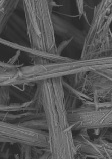
We image the surfaces and transects of samples using electron microscopy. The Stanford Mineral and Microchemical Analysis Facility offers a JEOL JXA-8230 “SuperProbe” with both EDS and WDS capabilities, and a brand new JSM-IT500HR InTouchScopeTM Scanning Electron Microscope with EDS for high resolution images. Contact Dale Burns for more details.
3D Imaging

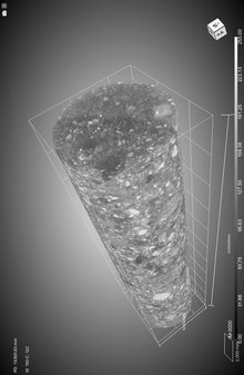
We use a Zeiss Xradia 520 Versa X-ray CT to acquire 3d images of rocks and geomaterials taken through various processes. You can learn more about the system available at the Stanford Nano Shared Facilities (SNSF) from Arturas Vailionis.
Chemical Analysis

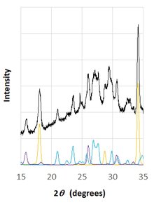
Techniques including XRD, XRF, ICP-MS/OES, FTIR and others are available on campus at the Environmental Measurement Facility (EMF) and Soft & Hybrid Materials Facility (SMF).
Mechanical Testing

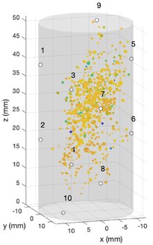
We utilize equipment in the Blume Earthquake Engineering Center for unconfined compression tests on rocks and geomaterials. Please contact Kyle Douglas for details on their many loading frames. We have used their MTS Criterion Series 40 for Brazilian split tests and uniaxial failure. Additionally, we constructed a coreholder assembly that affixes radial/axial strain gauges and 10 piezoelectric sensors to the sample, the latter of which can be used to measure acoustic emissions during deformation and ultimate mechanical failure.
